

Biology · cell structure
Animal cell 3D model with labels
Look inside a cutaway animal cell, learn what each organelle does, and use it for a school project.
By the Kenerate AI teamUpdated 2 min read
See the plant cell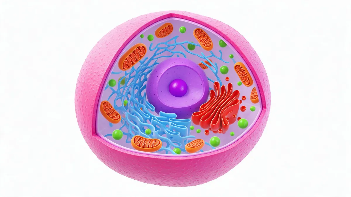
Quick answer
Updated
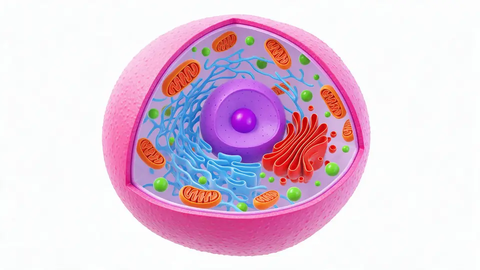
An animal cell is a eukaryotic cell surrounded by a flexible cell membrane, with no cell wall and no chloroplasts. Inside are a nucleus, mitochondria that release energy, endoplasmic reticulum, a Golgi apparatus, lysosomes, centrioles and ribosomes. The 3D illustration below is simplified for learning and not to scale.
Key facts
- Outer layer
- Cell membrane only — no cell wall
- Inside
- Nucleus, mitochondria, ER, Golgi, lysosomes, centrioles, ribosomes
- Found in animal cells
- Centrioles, lysosomes
- Energy
- Mitochondria (ATP)
- Illustration
- Simplified, not to scale
On this page
Animal cell 3D model
Animal cell parts, labeled
| Part | What it is | What it does |
|---|---|---|
| Cell membrane | A thin, flexible lipid bilayer [1] | Controls what enters and leaves the cell |
| Nucleus | Holds DNA; the dark centre is the nucleolus [1] | Controls the cell; the nucleolus makes ribosome parts |
| Mitochondria | Double-membrane organelles with folded inner membranes [1] | Release energy as ATP |
| Rough ER | Membranes studded with ribosomes [1] | Make and fold proteins |
| Smooth ER | Membranes without ribosomes [1] | Make lipids, detoxify |
| Golgi apparatus | Stacked flattened sacs [1] | Sort, modify and package proteins |
| Lysosomes | Sacs of digestive enzymes [1] | Break down waste and worn-out parts |
| Centrioles | Pair of tube-shaped structures [1] | Help organise cell division |
Animal cell vs plant cell
| Animal cell | Plant cell | |
|---|---|---|
| Cell wall | No | Yes (cellulose) |
| Chloroplasts | No | Yes |
| Vacuole | Small, if any | One large central vacuole |
| Centrioles | Present | Usually absent |
| Lysosomes | Common | Rare |
How to make an animal cell 3D model for a school project
- Build it from clay, a halved foam ball or cardboard — one colour per organelle — or print one.
- Make a 3D model with AI in the Kenerate 3D Studio from a short description, then print it.
- Add labels with small flags or a printed key.
- Mention that it's simplified: real cells are microscopic and contain far more of each organelle.
How it works
How it works in the app
Step 1
Choose text, photo or multi-view
Open 3D Studio and pick how to start: describe an object, upload a photo, or add several views.
Step 2
Pick a 3D engine
Open the model card to choose the engine and version that fits your object.
Step 3
Describe the object
Write one object on a plain background, then set textures, PBR materials and quality.
Step 4
Click Build 3D model
Press Build 3D model to start — the textured model appears in your gallery.
See it in the app

Open 3D Studio and pick how to start: describe an object, upload a photo, or add several views.


FAQ
Animal cell 3D model: questions
What are the main parts of an animal cell?
Cell membrane, cytoplasm, nucleus, nucleolus, mitochondria, rough and smooth ER, Golgi apparatus, lysosomes, centrioles, ribosomes and vesicles.
Link to this answerDo animal cells have a cell wall?
No — only a flexible cell membrane. Plant cells have both.
Link to this answerHow do I make a 3D animal cell model?
Build one from craft materials, or make one in a 3D app or with AI and print it.
Link to this answerWhat should the labels on an animal cell 3D model show?
Label at least the cell membrane, cytoplasm, nucleus, nucleolus, mitochondria, rough and smooth ER, Golgi apparatus, lysosomes, centrioles and ribosomes, each with a one-line job — the parts table above gives you the wording. Add a note that the model is simplified and not to scale.
Link to this answerCan I 3D print a cell model?
Yes — make one in a 3D app or with AI, export STL and print it in PLA; paint each organelle a different colour.
Link to this answerIs this illustration accurate?
It shows the main structures and roughly where they sit, simplified and not to scale.
Link to this answerSee the plant cell
Compare it with the plant cell 3D model — or make your own model with AI.